
Environmental Mastitis
Environmental mastitis is mostly caused by unsanitary bedding material that contain bacteria such as E. coli and Streptococcus uberis2 . Environmental mastitis can be prevented, at least in part, by applying pre-dip before milking6 and removing manure from bedding.
Contagious Mastitis
Contagious mastitis is exposed through milking protocols not being followed and unsanitary milking equipment from an infected cow that was milked previously5 . Common contagious bacteria include Staphylococcus aureus and Streptococcus agalactiae which are Gram-positive species. The best way to prevent contagious mastitis is to use a teat disinfectant after milking5 and using correctly cleaning milking equipment.
On-Farm Bacterial Culture
Being the gold standard for identification of mastitis pathogens, bacterial culture is an on-farm method that can provide results in 24 h.
Materials needed for culturing:
- Incubator set at 37℃ (98.6ºF)
- Culture plates (Tri-plate, Bi-plate, blood agar, MacConkey etc.)
- Disposable gloves
- Milk sample vials
- Sterile cotton swabs
- Cotton swabs soaked in 70% ethyl alcohol for teat disinfection
- Cooler with ice (if transporting milk samples to lab)
- Permeant marker for labeling
Next, a sterile cotton swab should be submerged in the milk sample until the swab is saturated with milk. Then apply the swab to the culture plate by the pattern shown below (Figure 1), and re-dip the swab in the milk for each of the three sections. After the milk samples are applied to the plate, label plates with cow ID and date. Then place the plates lid down in the incubator at 37℃ for 24 h7 .

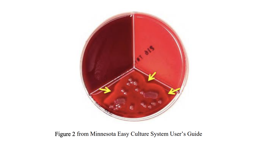
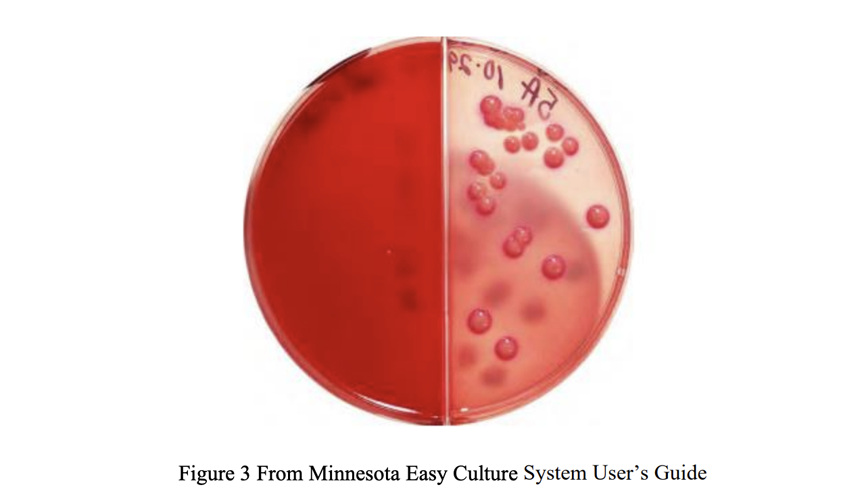
Interpreting the tri-plates results are different for each agar. Factor agar selectively grows streptococci spp, while Focus agar selectively grow staphylococcus spp1 . MacConkey agar is designed to grow Gram-negative bacteria. The simple diagnosis can tell the producers if treatment with antibiotics is necessary. Figure 2 represents further interpretation of Staphylococcus aureus with growth on the Factor agar. Figure 3 represents growth of E. coli which grows on MacConkey agar because this bacterium is Gram-negative. If plates contain three or more bacteria, then it is considered contaminated, and plating should be completed again6 . If the plate results in no growth, then that can be considered a true negative (no infection) or there was an error in the collection7 . If more than 40% of results are no growth, it is best to consult your veterinarian about diagnosis and treatment protocols7 . Bacteria such as Mycoplasma species cannot be cultured, so further methods are needed for identification such as quantitative polymerase chain reaction (qPCR) or a cell stain.
Conclusion
On-farm bacterial culture provides benefits to the producer to overall reduce mastitis cases. This is useful in providing the producer with information
References
- Lago, A., S. M. Godden, R. Bey, P. L. Ruegg, and K. Leslie. 2011a. The selective treatment of clinical mastitis based on on-farm culture results: I. Effects on antibiotic use, milk withholding time, and short-term clinical and bacteriological outcomes. J. Dairy Sci. 94:4441–4456.
- Derakhshani, H., Fehr, K.B., Sepheri, S., Francoz, D., De Buck, J., Barkema, H.W., Plaizier, J.C., Khafipour, E. 2018. Invited review: Microbiota of the bovine udder: Contributing factors and potential implications for udder health and mastitis susceptibility. Journal of Dairy Science. 10.3168/jds.2018-14860
- Liang, D., Arnold, L., Stowe, C., Harmon, R., Bewley, J. 2017. Estimating US dairy clinical disease costs with a stochastic simulation model. Journal of Dairy Science. 10.3168/jds.2016- 11565
- Barkema, H., Schukken, Y., Zadocks, R. 2006. Invited review: The role of cow, pathogen, and treatment regimen in the therapeutic success of bovine Staphylococcus aureus mastitis, Journal of dairy science. 10.3168/jds.S0022-0302(06)72256-1
- Hogan, J.S., Martin, M. 2016. Current concepts of bovine mastitis. Fifth edition. National Mastitis Council.
- Enger, B., Fox, L., Gay, J., Johnson, K. 2015. Reduction of teat skin mastitis pathogen loads: Differences between strains, dips, and contact. Journal of Dairy Science. 10.3168/jds.2014-8622
- University of Minnesota Laboratory for Udder Health, 2016. Minnesota Easy Culture System User’s Guide.



Be the first to comment